More from daniel yanez




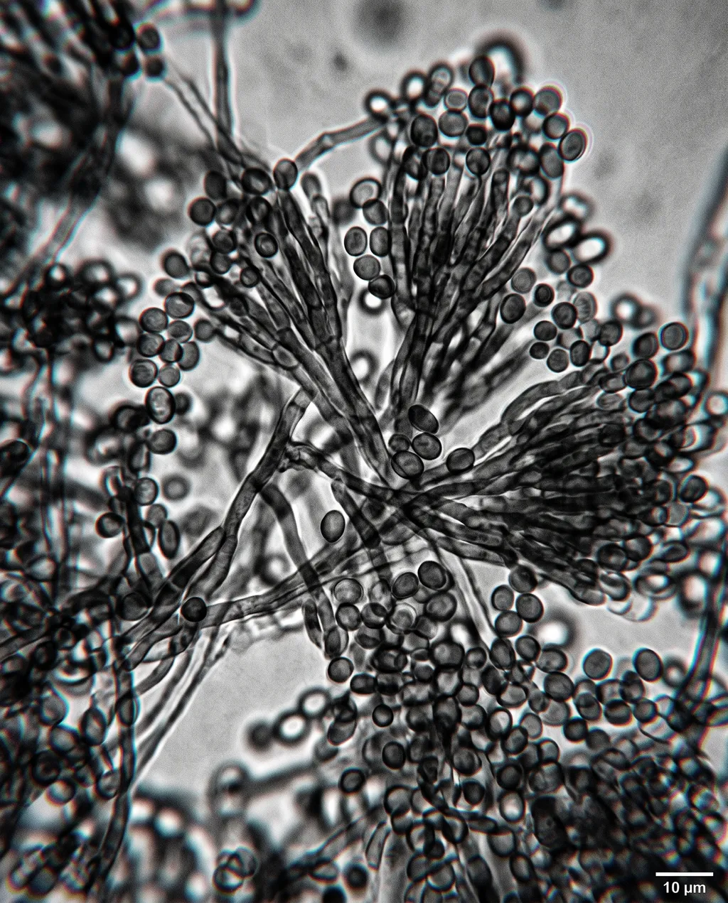
Microscopic view of Aspergillus fungus colony
Image by Nano Studio Pro · daniel yanez — free under CC BY 4.0
This high-magnification brightfield microscopy image reveals the intricate structure of an Aspergillus fungal colony. Centered in the frame is a prominent conidial head, showing the classic radiate arrangement of phialides and extending chains of conidia spores . The conidiophore stalk provides the structural support, rising from a dense, tangled mycelial network that spreads across the lower portion of the slide. Individual conidia spores are clearly visible, dotting the field of view with their spherical shapes and varying focal depths. The lighting is characteristic of phase-contrast or brightfield microscopy, emphasizing the edges of the cellular structures against a slightly luminous, grainy background. A 10 μm scale bar is positioned in the bottom right corner for size reference. The monochromatic aesthetic highlights the complex architectural morphology of the fungal reproductive structures.
More from daniel yanez