More from daniel yanez


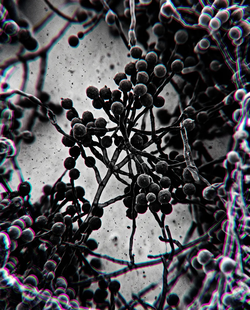
Microscopic view of fungal mycelium network
Image by Nano Studio Pro · daniel yanez — free under CC BY 4.0
This is a high-magnification scanning electron micrograph capturing the intricate and complex structure of a fungal mycelium network. The image presents a monochromatic, high-contrast view of interwoven filaments, known as hyphae, which branch out in a dense, web-like formation. Scattered across the delicate threads are numerous spherical fruiting bodies or sporangia, clustered together like heavy, dark berries on a vine. The lighting appears artificial, likely generated by the scanning electron microscope process, casting sharp highlights on the rounded surfaces and deep shadows within the web of fibers. The image has been processed with a subtle chromatic aberration effect, visible as slight blue and magenta fringes along the edges of the structures, adding a psychedelic or glitch-art aesthetic to an otherwise scientific specimen. The composition is highly detailed, highlighting the repetitive and chaotic beauty of organic growth patterns at a microscopic scale, rendered in a striking black, white, and grey palette.
More from daniel yanez


