More from daniel yanez


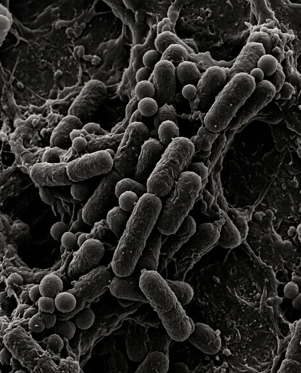
Scanning electron microscopy of mixed bacteria
Image by Nano Studio Pro · daniel yanez — free under CC BY 4.0
This grayscale scanning electron micrograph reveals a dense, complex microbial community organized into a biofilm. The visual field is dominated by a mixture of rod-shaped bacilli and spherical cocci, which are interconnected by a web-like extracellular polymeric substance (EPS) matrix . The lighting, characteristic of SEM, creates sharp highlights and deep shadows that emphasize the three-dimensional, pill-like morphology of the individual cells and the rounded, smooth surfaces of the cocci . The composition shows a high degree of structural heterogeneity, typical of bacterial biofilms attached to a substrate. The monochrome palette accentuates the textural nuances of the bacterial cell walls and the filamentous matrix bridging them. This image is a stark, scientific representation of microbial colonization, showcasing the varied growth patterns and density of the colony at high magnification.
More from daniel yanez


